Musculoskeletal ultrasound at EBP Studio Amsterdam: a valuable addition to physiotherapy diagnostics
Ultrasound, or musculoskeletal ultrasound (MSU), is an imaging technique that uses high-frequency sound waves to image structures in the body. The method is particularly suitable for soft tissues such as tendons, muscles, ligaments, nerves, and superficial joints. Thanks to its high resolution and the possibility of dynamic research, ultrasound has become an important diagnostic tool in orthopedics, rheumatology and sports medicine. At EBP Studio in Amsterdam South, we use this technique as an addition to regular physiotherapy research, with the aim of refining our diagnoses and better tailoring treatment programs to the individual patient.
What is Musculoskeletal Ultrasound?
MSK ultrasound uses high-frequency sound waves to form an image of structures such as tendons, muscles, ligaments, bursa, nerves, and superficial joint capsules. In contrast to X-ray testing, ultrasound does not involve radiation, making it a safe method that can be used repeatedly without risk to the patient. The research takes place in real-time, which means that dynamic movements of the joint or muscle groups are immediately visible on the screen.
But ultrasound also has limitations: certain structures are difficult or not visible at all. In this blog, we highlight what, according to the latest guidelines from the European Society of Musculoskeletal Radiology (ESSR), can and cannot be reliably imaged with ultrasound. In doing so, we refer only to literature from the 2018 ESSR Consensus Update.
Benefits and applicability of ultrasound
Ultrasound has a number of clear advantages over other imaging techniques:
- The research is not burdensome (no radiation).
- It can be deployed dynamically (the movement of structures can be assessed live).
- It is accessible, widely available and relatively cheap.
- There is immediate feedback, which is valuable for both diagnosis and guidance for injections.
In fact, in many cases, ultrasound is the preferred method. The ESSR works with a grading scale from 0 to 3 to indicate the extent to which ultrasound is suitable for a particular indication:
0: not indicated
1: indicated when other techniques are not possible
2: equivalent to other imaging
3: first choice
Ultrasound in physiotherapy is particularly suitable for imaging:
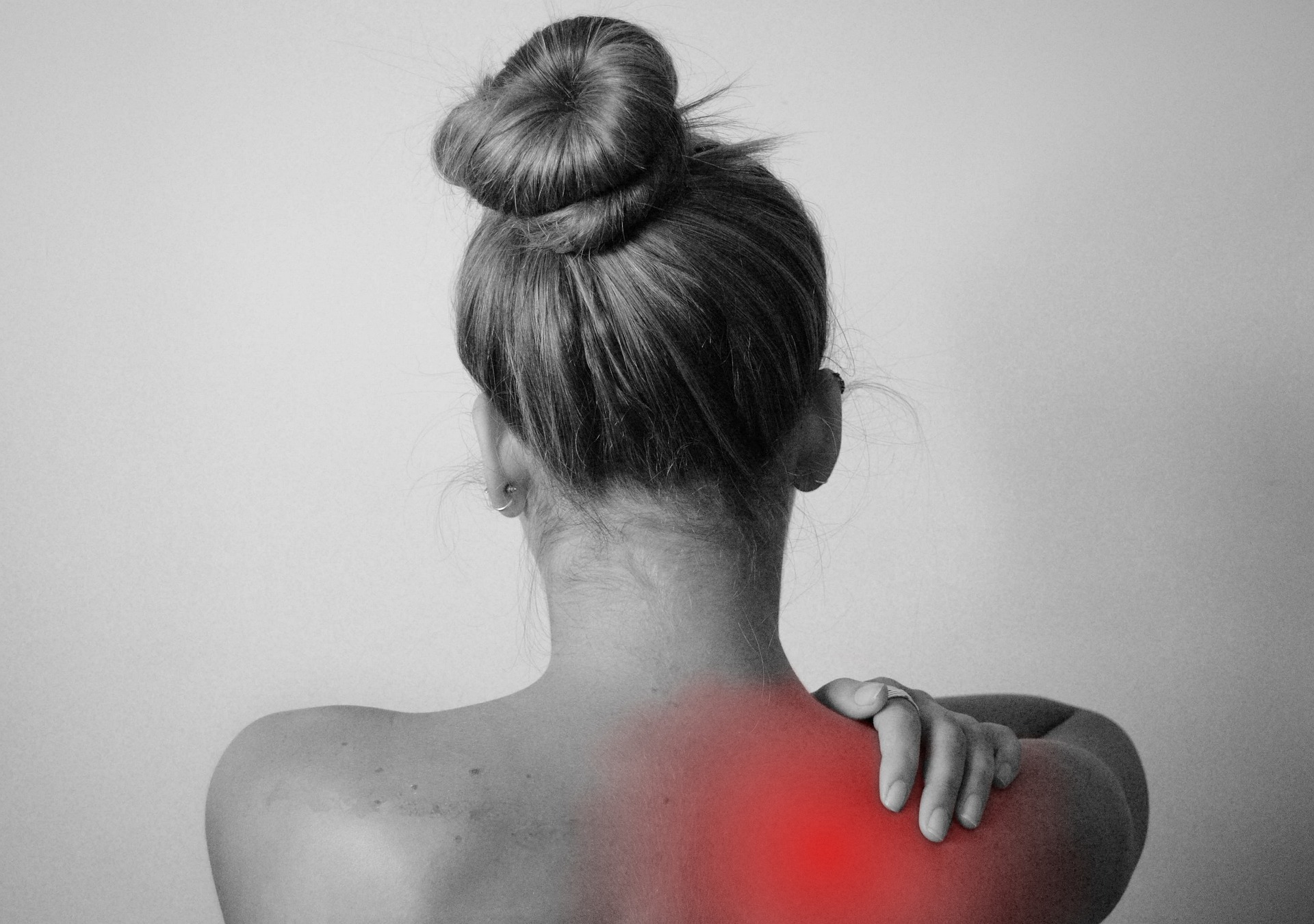
Shoulder:
Ultrasound is often the first choice for shoulder problems:
- Complete rotator cuff ruptures (grade A, consensus 3)
- Partial cuff ruptures and rotator cuff calcifications and tendon luxations of the long biceps tendon can be reliably imaged (grade A/B, consensus 2-3).
Elbow
The following structures can be easily evaluated with ultrasound:
- Lateral and medial epicondylitis (grade A/B, consensus 3)
- Triceps tendon, olecranon bursitis and synovitis (grade C/A, consensus 3)
- Ulnaropathies such as compression or subluxation of the ulnar nerve (grade B/C, consensus 3).
Wrist and hand
- Tenosynovitis, tendon ruptures, pulley lesions, trigger finger and ganglia are clearly visible (grade B/C, consensus 3).
- Carpal Tunnel Syndrome (CTS) is reliably detected with a consensus of 3 and an evidence level of B.
Hip
Ultrasound is very valuable for structures on the outside of the hip:
- Gluteal tendon ruptures and gluteal tendinopathy (Grade A, consensus 2)
- Snapping hip (extra-articular) (grade A, consensus 3)
- Trochanteric bursitis, hamstrings and Morel-Lavallée lesions (grade B/C, consensus 2-3).
Knee
- Patellar tendon and quadriceps ruptures, bursitis, Baker cysts and Osgood-Schlatter are clearly visible (grade A/C, consensus 3).
- The medial collateral band and semimembranosus/semitendinosus tendons are clearly visible (grade A/B, consensus 2-3).
Ankle and foot
Ultrasound is the first choice for:
- Achilles tendon lesions, plantar fasciitis, calcaneal bursitis, ligament injuries (such as the ATFL), and Morton neuromas (grade A/B, consensus 3).
- Plantar plate ruptures and ankle instability have been added to the list of indications with grade A and consensus 2.
Nerves
Various nerves can be reliably imaged:
- n. medianus (CTS), n. ulnaris, n.radialis, n. ischiadicus, n. cutaneus femoris lateralis (grade B/c, consensus 3).
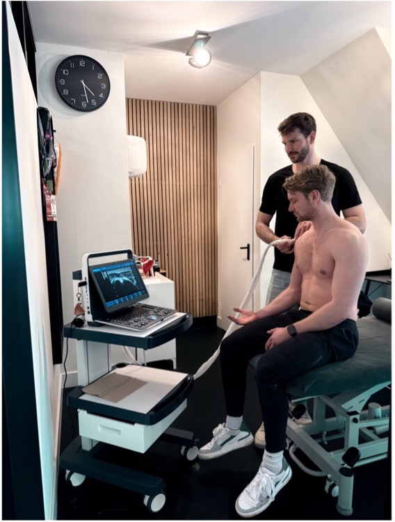
Ultrasound at EBP Studio
At EBP Studio, MSK ultrasound is performed by physiotherapist Julian Jaring, who specializes in examining complaints of the shoulder, elbow, hip, knee and hand. If a tendinopathy (e.g. in the shoulder or knee) or a partial muscle tear (such as a hamstring injury) is suspected, ultrasound can provide clarity about the severity and location of the injury.
The results of the ultrasound are discussed directly with the patient. This promotes insight into the nature of the complaint and increases the patient's involvement in the treatment process.
In addition, the ultrasound is regularly used for:
- Second opinions in case of long-term or misunderstood complaints.
- Monitoring recovery after an injury or surgery.
- Substantiation of referral to a doctor or specialist when indicated.
Limits of ultrasound
While MSK ultrasound can provide a lot of information, there are also clear limitations. Structures that lie deeper in the body — such as the hip joint, spine, or cartilage within joints — are often not clearly visible. This is because ultrasound sound waves cannot penetrate bone. As a result, structures that are behind bone, such as the meniscus or spinal canals, cannot be reliably imaged.
In case of doubt or suspicion of intraarticular damage (e.g. a torn cruciate ligament or labrum tear), the physiotherapist may decide to refer for additional examinations, such as an MRI.
Deeply located or intra-articular structures
- Intra-articular labrum tears of the hip and shoulder are not clearly visible (grade C or 0).
- Meniscal injuries in the knee: despite new publications, it remains recommended to be cautious. MRI remains the standard here (grade A, but consensus 0).
- Crucial ligaments (ACL/PCL) are usually not visible due to their location (grade A/B, consensus 0).
- Cartilage damage in the ankle and knee is limited (grade D, consensus 0 or 1).
Complex joint instability
- Dynamic instability of the glenohumeral joint (e.g. shoulder dislocations): ultrasound is not suitable for this (grade B, consensus 0).
Bone structures and deep fractures
- Intra-articular fractures and fractures of the pelvis, for example, are not easy to assess with ultrasound. X-ray or CT are indicated here (grade C, consensus 0-1).
- Osteochondritis dissecans, synovial tumors and plica syndrome are also outside the scope of ultrasound (grade C/D, consensus 0).
Pelvic physiotherapy and ultrasound
A special area of application within our practice is pelvic physiotherapy. Here, we use ultrasound to assess the pelvic floor muscles, for example after pregnancy or in case of complaints such as urinary incontinence or prolapse. The ultrasound makes it possible to objectively measure the state of tension, coordination and activity of the pelvic floor. This is important for determining the right time to return to exercise or resume certain physical activities.
Pelvic floor ultrasound offers insight into, among other things:
- The degree of activation during tension and relaxation
- The degree of diastasis after pregnancy
- Bladder contents
- Functional employability during movement
For many patients, it is pleasant and comforting to see what is happening in their bodies on screen. This increases confidence and motivation to keep practicing.
Why choose ultrasound at EBP Studio Amsterdam?
For patients seeking physical therapy in Amsterdam South (Rivierenbuurt), EBP Studio Amsterdam offers high-quality diagnostics combined with personal care. Ultrasound is a valuable addition to physiotherapy research because:
- It helps to make a more precise diagnosis
- It provides insight into the structure and function of soft tissues
- The recovery can be monitored objectively
- The patient is directly involved in the findings
Especially in the case of complex or long-term complaints, ultrasound can make the difference between general and customized treatment.


.jpg)
.jpg)


